

Breast cysts – definition and treatment

What is a fibroadenoma?
The lumps may be hard or flexible, and are usually painless. However, sometimes they grow so big that they are visible or cause breast pain. In such cases, the tumour is removed surgically. Fibroadenomas are relatively common in women. One in four women have more than one tumour, but after menopause the tumours regress and calcify.
A characteristic feature of fibroadenoma is that it moves under the fingers and has an irregular shape. It may be hard or flexible, and is usually painless. Fibroadenomas sometimes become enlarged with the passage of time and may grow to an extent that they are visible or cause breast pain. In such cases, the tumour is removed surgically. The tumour may also grow in size during pregnancy and breastfeeding. Fibroadenomas are relatively common in women. One in four women have more than one tumour, but after menopause the tumours have a tendency to regress and calcify.
Types of fibroadenomas
Simple fibroadenoma consists of glandular and fibrous tissue and is not dangerous.
In a complex fibroadenoma, there is a presence of other breast tissues apart from the glandular and fibrous tissues. Complex fibroadenomas increase the risk of breast cancer. Observations or histopathological examinations are recommended, especially for elderly women with genetic or family predispositions as they have a two-fold higher risk of developing breast cancer when compared to other women.
Giant fibroadenoma – a tumour more than 5 cm in diameter.
Juvenile fibroadenoma – a tumour occurring in adolescents.
Diagnosis and treatment of fibroadenomas
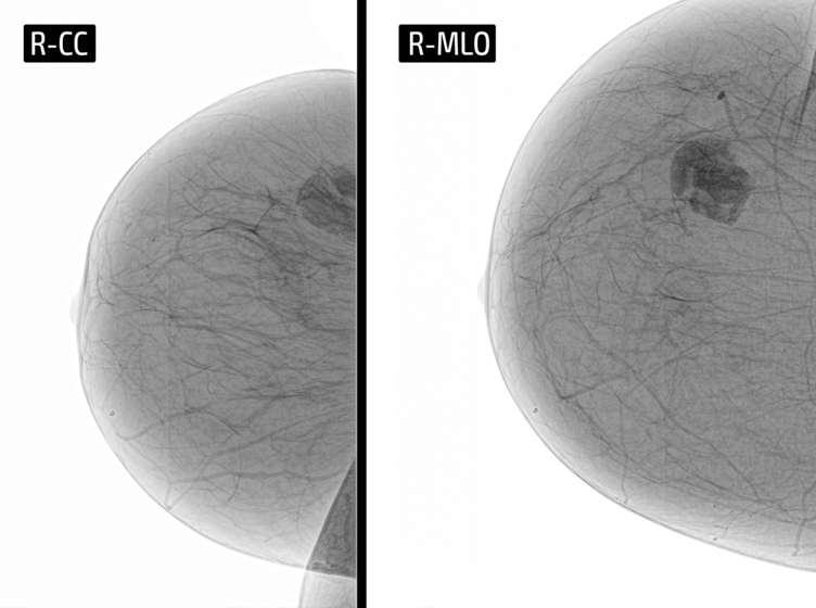
Depending on the patient's age, different diagnostic tests are performed. In young women under the age of 35, a palpation (touch test) and a breast ultrasound scan are performed. If the scans do not have any abnormalities, no further tests are required. However, the change should be observed. In women over 35 years of age, mammography is performed. On a mammogram, fibroadenoma appears as a mass with smooth, rounded edges which are distinct from the surrounding tissue. To further evaluate the fibroadenoma, a fine-needle or a core-needle biopsy may be performed.
Now it is possible to self-examine the breast at home, thanks to the innovative Braster device.
Fibroadenoma, fibroma, adenoma and hormonal contraception
In a majority of women with such tumours, fibroadenomas develop slowly. However, pregnancy or hormonal therapy, as well as contraceptive pills containing oestrogen are conducive to their growth and may induce proliferation.
Removal of fibroadenomas
Fibroadenomas are removed if there are histopathological abnormalities. In other cases, it is recommended to remove the tumour if it is larger than 4 cm, or if it grows very quickly. The tumours should also be removed if they cause pain or affect the symmetry of the breast. Surgical procedures are often performed for aesthetic reasons. Unfortunately, removal of the tumour does not guarantee that it will not reoccur. Therefore, it is of utmost importance to self-examine the breast regularly and do an ultrasound or mammography every year, even after the removal of fibroadenomas.
Zostaw swój numer, oddzwonimy i odpowiemy na Twoje pytania
I consent to the use of telecommunication end devices by BRASTER S.A. for commercial purposes and direct marketing, pursuant to Article 172 of the Act of July 16, 2004 – Telecommunications Law and the Act of July 18, 2002 on electronic provision of services. In particular, I consent to receiving incoming phone calls from BRASTER S.A. I declare that I have been informed that the above consent is voluntary and may be revoked at any time.
Strona http://braster.eu/nl realizuje sprzedaż jedynie na terenie Holandii. Jeżeli chcesz zrealizować zamówienie na terenie Polski przejdź na http://braster.eu/pl
I consent to the use of telecommunication end devices by BRASTER S.A. for commercial purposes and direct marketing, pursuant to Article 172 of the Act of July 16, 2004 – Telecommunications Law and the Act of July 18, 2002 on electronic provision of services. In particular, I consent to receiving incoming phone calls from BRASTER S.A. I declare that I have been informed that the above consent is voluntary and may be revoked at any time.
Your message was sent.
We will reply as soon as possible.
Raty PayU to możliwość zapłacenia za zakupy w ratach (od 3 do nawet 36)
Sign In
Create New Account