

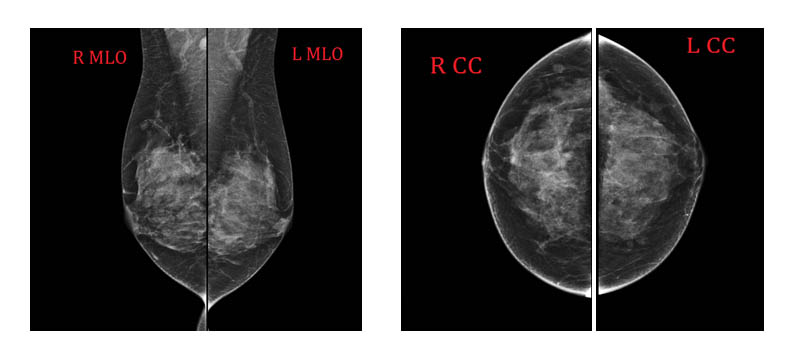
40 year old woman, undergone mammography which revealed dense glandular breast composition. Oval, well circumscribed foci up to 10mm in diameter were seen, no suspcious mass or clusters of calcifications was found, qualified patient for an additional ultrasound evaluation,
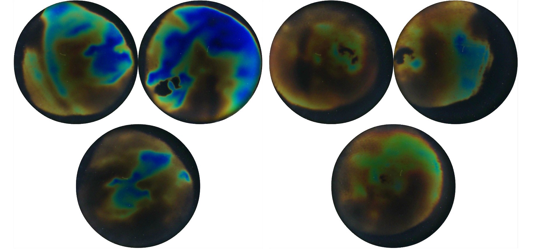
BI-RADS 0. On ultrasonography several simple cyst were noted in both breast. In the right breast at the 12 o’clock axis irregular hypoechoic lesion 33x18mm was identified, suspicious of maliganancy. BI-RADS 5. Thermography showed irregular hyperthermy localised in the centre of right breast, on the border of the upper quadrants. Moreover scattered small hyperthermic spots are seen in both breast.
Core biopsy confirmed ductal carcinoma, and the fine needle biopsy confirmed cancerous cells in the axillary lymph nodes. Due to the dense breast structure, breast cancer in this case was not visible on mammography. It was revealed in the ultrasound examination as an irregular hypoechogenic change, and on thermographic images as an asymmetric structure with a higher thermal degree within the analogous right breast quadrant.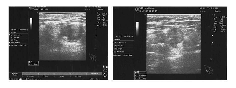
Sign In
Create New Account