

Breast calcifications – their definitions and types
Phyllodes tumors of the breast may be both benign and malignant. Most are benign; however, even histologically benign phyllodes tumors can be locally malignant. Phyllodes tumors occur rarely – they account for less than 1% of all breast tumors. Women aged 45–49 are at the highest risk. However, sporadically, teenagers may also suffer from this disease. Phyllodes tumors are located in the stroma of the breast tissue. They can be tiny – about 3–4 cm in diameter – but most of them grow very large, expanding the breast – they can grow as big as 15 cm. According to the literature, the change is palpable in 80% of patients with phyllodes tumors, and in 20% the pathology is not detected until discovery by routine screening tests. In cases with palpable phyllodes tumors, a rapid increase in the size of the tumor over a short time is observed. In most cases, the removal of the tumor means complete recovery; however, some cases of recurrence have also been observed.
Symptoms
In most cases, a distortion of the skin and nipple can be observed due to the pressure of the tumor. The symptoms are quite non-specific. There is no pain; therefore, it is important to pay attention to any thickenings of breast tissue that occur, elevated temperature in one breast when compared to the other, the appearance of an area of redness on the skin of the breast, or skin ulceration above the change.
Types
It is difficult to determine the difference between benign and malignant changes. That is why phyllodes tumors are divided into three categories:p>
benign - doubles its weight in approximately 116 days, the margins of the benign tumors extend into the surrounding tissues, there is no infiltration of surrounding tissues. When resected, the risk of recurrence is very low;
limited malignancy - related to a 25% risk of recurrence and 5% risk of metastases;
malignant - doubles its weight in approximately 36 days, its borders significantly infiltrate surrounding tissues, similar to some breast cancers. Sometimes the epithelial elements can grow; in exceptional cases, this growth turns into cancer. Such a transition occurs rarely. This is probably because the rapid growth of a tumor leads to it manifesting and, as a result, being removed before such a transition takes place. In the case of this type of tumor, the risk of local recurrence and metastases are high, about 25%. Therefore, it is recommended to resect the tumor together with a large margin. These tumors rarely metastasize via the lymphatic vessels to the axillary lymph nodes, but they may metastasize via the blood to the lungs, liver and other organs located further afield. They can result in the patient’s death.
Diagnosis
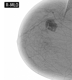
Phyllodes tumors can only be diagnosed with the use of a guided biopsy, in which puncturing and sampling are performed under the control of a mammograph or an ultrasound machine. If the guided biopsy does not result in the full identification of the cancer, an open surgery biopsy is performed. Using local anesthesia and under the control of a mammograph or an ultrasound machine, the doctor removes marked changes together with some parts of surrounding tissues. After microscopic examination of the tissue sample, the type and size of the tumor are determined and the doctor decides on the treatment method.
Treatment
Only treatment through surgery is possible, since the tumor is not sensitive to radiotherapy or chemotherapy. Benign changes are removed together with a small margin of the surrounding tissues. More serious cases require mastectomy; i.e., amputation of the whole mammary gland together with the nipple. However, axillary lymph nodes are not removed, because the tumor metastasizes via the blood, not lymphatic vessels.
[1] V. Kumar, R. S. Cotran, S. L. Robbins; Patologia [Pathology]; Urban & Partner 2005.
[2] K. Hemminki, C. Granström; Morphological types of breast cancer in family members and multiple primary tumours: Is morphology genetically determined? Breast Cancer Res 2002; 4: R7.
[3] T. A. Stavros; Ultrasonografia Piersi [Breast ultrasonography]; Radiology Imaging Associates, Polish Edition, Warszawa 2007.
Zostaw swój numer, oddzwonimy i odpowiemy na Twoje pytania
I consent to the use of telecommunication end devices by BRASTER S.A. for commercial purposes and direct marketing, pursuant to Article 172 of the Act of July 16, 2004 – Telecommunications Law and the Act of July 18, 2002 on electronic provision of services. In particular, I consent to receiving incoming phone calls from BRASTER S.A. I declare that I have been informed that the above consent is voluntary and may be revoked at any time.
Strona http://braster.eu/nl realizuje sprzedaż jedynie na terenie Holandii. Jeżeli chcesz zrealizować zamówienie na terenie Polski przejdź na http://braster.eu/pl
I consent to the use of telecommunication end devices by BRASTER S.A. for commercial purposes and direct marketing, pursuant to Article 172 of the Act of July 16, 2004 – Telecommunications Law and the Act of July 18, 2002 on electronic provision of services. In particular, I consent to receiving incoming phone calls from BRASTER S.A. I declare that I have been informed that the above consent is voluntary and may be revoked at any time.
Your message was sent.
We will reply as soon as possible.
Raty PayU to możliwość zapłacenia za zakupy w ratach (od 3 do nawet 36)
Sign In
Create New Account